New study examines cost-effective imaging that protects patients and health care workers from COVID-19 exposure
By Christy Janssens
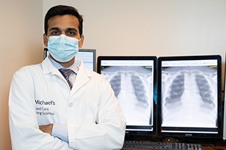
Dr. Shobhit Mathur
How can we keep COVID-19 patients and their care teams safe while taking chest scans to monitor their wellbeing?
One group of researchers at St. Michael’s Hospital studied an innovative technique to do just that. A study led by Dr. Shobhit Mathur, a physician at St. Michael’s, examined a new radiology process for obtaining chest images through glass doors of isolation rooms. This technique helps to avoid exposure to the virus, while providing clinicians the information they need to treat patients.
Different teams came together to swiftly and safely adopt this technique. This study’s findings were recently published in Investigative Radiology and Canadian Association of Radiologists Journal.
We sat down with Dr. Mathur to learn more about this work.
Q: What is this new technique and how does it differ from conventional imaging techniques?
A: Chest radiograph is a low-cost and widely available medical imaging tool commonly used to detect chest problems in patients with possible and confirmed COVID-19 infection. In conventional bedside radiography, two technologists must enter a patient’s isolation room with a machine to perform the imaging. This can increase the chances of exposure to a virus. Of course, when entering a COVID-19 patient’s room, both technologists also need to put on personal protective equipment (PPE) during the procedure and clean the machine afterwards.
Chest radiography through glass, however, is a novel technique that allows imaging through the protection of a glass wall. In this scenario, only one of the two technologists is required to enter the isolation room, while the other operates the machine from outside the room. While this technique has many benefits and was suggested by the Radiological Society of North America during the pandemic, no scientific evidence existed about the best way to use it. We believe that this is an important factor limiting its widespread adoption.
Q: What are the benefits of this technique?
A: This technique limits health-care personnel and patient exposure to COVID-19, conserves PPE (only one technologist needs to wear PPE) and decontamination resources, and reduces equipment downtime by eliminating the need to clean the machine after every use.
All of this enhances the protection of the patient and their care team, increases hospital efficiency, and reduces health-care costs.
Q: What were your key findings?
A: Our research project evaluated the feasibility, image quality and radiation safety of this technique in non-clinical and clinical settings.
Radiographic image quality is a complex interplay of several technical and patient factors. Through our research, we showed how technical parameters can be optimized to obtain good quality images while ensuring the least possible radiation to patients.
We also found minor backscatter radiation, a small amount of radiation that deflects from the glass and goes the opposite way of the target, which suggests that the technologist outside the room should use lead shielding or maintain appropriate distance from the glass door. This does not affect the patient being imaged.
Our research also found that we could save an estimated $50,000 annually in our Emergency Department by adopting this technique.
Q: What is next for this research?
A: We hope that our experience and results will help other hospitals adopt this technique. Our next step is to enhance collaboration with the industry to make this technique more user friendly. Looking ahead into the future, the ease and benefits of implementing this technique make a strong argument for its continuation in the post-COVID-19 era. This technique has the potential to prevent infection transmission while caring for patients isolated for other infectious illnesses requiring similar precautions.
This work was made possible through the collaboration of many different teams at St. Michael’s, including: the Radiology Department (Archana Rai, Darren Liu, Noah Ditkofsky, Djeven Deva, Tim Dowdell, Kate MacGregor, Bryce Hunt, Margaret Dubrawski, David Bristow, David Nelson); the Emergency Department (Alun Ackery); Procurement (Mark Linsao); and the Planning/Development department (Dominic Gascon). A faculty at Michener Institute, Alex Gontar, also worked with the St. Michael’s researchers to support this project.
About St. Michael’s Hospital
St. Michael’s Hospital provides compassionate care to all who enter its doors. The hospital also provides outstanding medical education to future health care professionals in more than 27 academic disciplines. Critical care and trauma, heart disease, neurosurgery, diabetes, cancer care, care of the homeless and global health are among the Hospital’s recognized areas of expertise. Through the Keenan Research Centre and the Li Ka Shing International Healthcare Education Centre, which make up the Li Ka Shing Knowledge Institute, research and education at St. Michael’s Hospital are recognized and make an impact around the world. Founded in 1892, the hospital is fully affiliated with the University of Toronto.
About Unity Health Toronto
Unity Health Toronto, comprised of Providence Healthcare, St. Joseph’s Health Centre and St. Michael’s Hospital, works to advance the health of everyone in our urban communities and beyond. Our health network serves patients, residents and clients across the full spectrum of care, spanning primary care, secondary community care, tertiary and quaternary care services to post-acute through rehabilitation, palliative care and long-term care, while investing in world-class research and education. For more information, visit www.unityhealth.to.