Use of simulated medical images improves accuracy of computers with AI
By James Wysotski
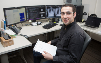
Artificial intelligence expert Hojjat Salehinejad compares real medical images with simulations created by computers with AI. (Photo by Katie Cooper)
Radiologists in the Medical Imaging Department might get a new assistant in the coming years that never sleeps or leaves the hospital.
In April 2016, Drs. Tim Dowdell, Joe Barfett and Errol Colak – all radiologists – created the Machine Intelligence in Medicine Lab to teach computers with artificial intelligence (AI) how to interpret medical images.
Computing systems with AI have been trained to think and learn like human brains. These systems progressively improve their performance on tasks by creating associations between data. The bigger the datasets, the better they learn. Once they’ve learned enough to demonstrate cognitive-like functions such as problem solving, they’re deemed to have AI.
Embracing AI could help radiologists improve quality and reduce errors, said Dr. Barfett. While medical mistakes are not frequent, they happen. Cancers occasionally get missed.
“With these AI tools, it’s very realistic to think that in the next five years no one will ever have a lung nodule accidentally missed on a chest X-ray,” said Dr. Barfett. “AI can make such instances go from rare to exceedingly rare.”
At present, he said there are limitations to what AI can do. For example, it can’t problem-solve to the degree that a human can. After flagging a potential lung nodule on an X-ray, it cannot ask why it’s there and start looking for the cause of the problem. That’s where a radiologist is needed.
However, adopting AI has other advantages.
“I suspect that AI can detect cancers that a human cannot because of how well it can sense subtleties on X-rays,” said Dr. Barfett.
Plus, everything gets interpreted in real time, thereby speeding up workflows and reducing wait times for patients.
“Eventually it will be just the AI doing the screening. It’s not the present, but there’s no question to me that it’s the future.” – Dr. Joseph Barfett |
Having computers flag potentially abnormal cases would also be a huge savings to the health-care system since it would allow radiologists to spend their time on more complicated, patient-care oriented problems, said Dr. Barfett.
Soon after forming the MIMLab, the three radiologists recruited AI expert Hojjat Salehinejad, a PHD student in the Electrical and Computer Engineering Department of the University of Toronto, who Dr. Barfett said is now the driving force of their research.
Together they have overcome some early stumbling blocks – the team discovered it could not sufficiently train AI algorithms to interpret X-rays using just hospital databases because the datasets were imbalanced, said Dr. Barfett. While the databases had numerous examples of common ailments, there were too few of the rarer conditions that also tended to be more life-threatening.
A unique solution was put in place and instead of relying solely on real medical images, the team augmented its database by programming AI algorithms to create computer-generated chest X-rays. Enough images of rare conditions were created, that when combined with the real ones it gave the team exactly what it needed to teach a machine how to spot conditions on a very broad spectrum – including those rare cases that could mean the difference between life and death for a patient.
By including the computer-generated images, Dr. Barfett said the computers’ ability to accurately interpret X-rays of common diseases improved by 20 per cent. For rarer conditions such as pneumothorax (a collapsed lung), the improvement was about 40 per cent.
With each step forward in its AI research, Dr. Barfett said the MIMLab helps determine the future of radiology.
“We’re trying to channel our efforts into things that have immediate impact,” said Dr. Barfett. “We can direct the scientific discourse toward clinical questions that we know could lead to significant improvements for patients in the next five years, even if that means giving away the AI innovations we create.”
About St. Michael’s Hospital
St. Michael’s Hospital provides compassionate care to all who enter its doors. The hospital also provides outstanding medical education to future health care professionals in more than 29 academic disciplines. Critical care and trauma, heart disease, neurosurgery, diabetes, cancer care, care of the homeless and global health are among the Hospital’s recognized areas of expertise. Through the Keenan Research Centre and the Li Ka Shing International Healthcare Education Centre, which make up the Li Ka Shing Knowledge Institute, research and education at St. Michael’s Hospital are recognized and make an impact around the world. Founded in 1892, the hospital is fully affiliated with the University of Toronto.
St. Michael’s Hospital with Providence Healthcare and St. Joseph’s Health Centre now operate under one corporate entity as of August 1, 2017. United, the three organizations serve patients, residents and clients across the full spectrum of care, spanning primary care, secondary community care, tertiary and quaternary care services to post-acute through rehabilitation, palliative care and long-term care, while investing in world-class research and education.