Retinal unit eyes even better safety with new technology
By James Wysotski
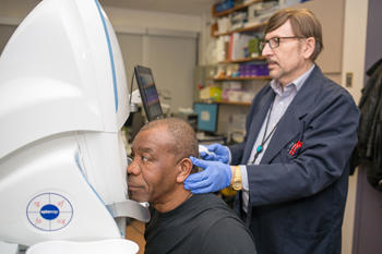
Ophthalmic technician Vladimir Evlampiev takes pictures of Eustace Nembhard’s right eye using wide-angle angiography. (Photo by Yuri Markarov)
There’s no limit to better, even for the largest retina unit in the country.
The Eye Clinic at St. Michael’s already uses a diagnostic machine that is unique among Canadian hospitals and soon it will get another kind that’s even better, said Ophthalmologist-in-Chief Dr. David Wong.
Since July 2016, the clinic has been taking pictures of blood vessels at the back of the eye using wide-angle angiography, which involves injecting fluorescein dye into a patient’s arm that flows to blood vessels in the eyes and allows them to be seen more easily by doctors’ cameras. Typically, angiography machines offer a narrow field of view, but the hospital’s wide-angle unit allows St. Michael’s physicians to see more than anyone else in Canada, said Dr. Wong. By injecting a second dye, indocyanine green, the field of view expands to the retina’s periphery.
“We’re finding diseases in the eye that we couldn’t see in the past,” said Dr. Wong. “Now, we can actually view what we had always assumed to be true.”
Dr. Wong said the clinic sees a lot of people with diabetes, but also vascular diseases such as hypertension and eye strokes. There’s also talk of detecting Alzheimer’s disease earlier through thinning of the retina. The clinic also deals with uveitis – inflammation of the eye – and Dr. Wong said that once inflammation is found in the eye, it’s usually somewhere else in the body, often in the form of arthritis, colitis or Crohn’s disease.
“We’re defining what’s normal and, ultimately, what’s abnormal,” said Dr. Wong. “And it’s changed the way we do patient care.”
“As an academic unit, if we change our thinking, we change everybody else’s because we are the leaders.” – Dr. David Wong, Ophthalmologist-in-Chief |
Diagnostics used to be somewhat qualitative, said Dr. Wong, but the new machines offer enough scope and refinement to make accurate measurements down to five one-thousandths of a millimeter. As a result, therapies are safer since the effectiveness of drug treatments can be better evaluated.
As good as wide-angle, fluorescein angiography is, the future is digital.
Instead of injecting dye to see the eye’s blood vessels, new optical coherence tomography, or OCT, machines use lasers to obtain cross-sectional images non-invasively. The technology scans so quickly that “we can actually see the retina in living detail,” said Dr. Wong.
Like many clinics, St. Michael’s already owns OCT machines. However, Dr. Wong said that by fall, the hospital will be the first in Canada to get the next generation of the technology, swept source OCT, which provides a wide-angle view and even greater resolution.
The new machine has other benefits. While the technology with the dye requires 20 minutes with a nurse and a photographer, OCT needs one technician for just two minutes, meaning that more patients can be seen at less of a cost to the health-care system. And since two per cent of patients have an allergy to the fluorescein dye, OCT’s non-invasiveness improves safety.
“We’re going from a safe technology to an ultra-safe one,” Dr. Wong said.
About St. Michael’s Hospital
St. Michael’s Hospital provides compassionate care to all who enter its doors. The hospital also provides outstanding medical education to future health care professionals in 27 academic disciplines. Critical care and trauma, heart disease, neurosurgery, diabetes, cancer care, care of the homeless and global health are among the hospital’s recognized areas of expertise. Through the Keenan Research Centre and the Li Ka Shing International Healthcare Education Centre, which make up the Li Ka Shing Knowledge Institute, research and education at St. Michael’s Hospital are recognized and make an impact around the world. Founded in 1892, the hospital is fully affiliated with the University of Toronto.